Development of a luminescent substance with nanoparticles that allows more accurate and safe viewing of different parts of the body
A research team from the Institute of Materials Science of Seville (a CSIC -Spanish National Research Council- and University of Seville joint centre) has designed a fluid that works like a luminous ink to obtain very sharp images of damaged tissues, organs and cartilages in diagnostic tests. This new compound, still in the laboratory phase, reduces adverse effects on the human body because it allows lower amounts to be injected and the dose to be targeted only at the affected area.
A luminescent fluid made up of nanoparticles that allows more accurate observation of different areas of the organism has been developed by a research team from the Colloidal Materials group of the Institute of Materials Science of Seville (a CSIC-University of Seville joint centre) in collaboration with the Institute of Materials Science of Aragon, the Andalusian Centre of Medicine and Biotechnology (BIONAND) and the Spanish National Accelerator Centre (CNA). This substance, still in the laboratory phase, offers a number of advantages over those currently used: it reduces the adverse effects on the body, uses lower doses than usual, allows the compound to be targeted only at the affected area that needs to be visualized, and is very versatile, since the same substance can be used for different diagnostic tests.
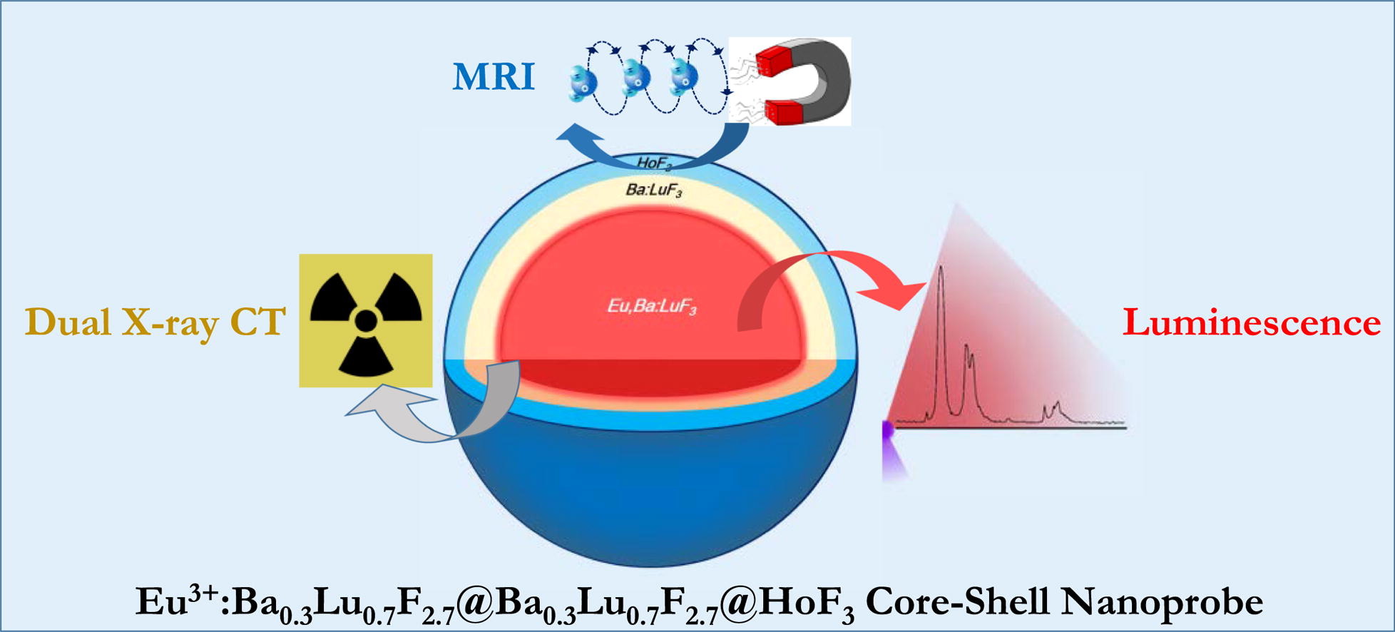
The nanoparticles consist of a nucleus, an intermediate coating layer and an outer shell.
Substances that serve to ‘light up’ and allow better observation of damaged organs, tissues or cartilages are called contrast agents. These act like an ink to better define the parts of the anatomy that need to be viewed through diagnostic imaging techniques. These substances are used in various tests that provide three-dimensional images of organs and tissues of the human body: such as computed tomography (CT), which is used to observe damaged organs through X-rays, as in a conventional X-ray scan; or such as magnetic resonance imaging (MRI), a diagnostic test based on a machine that uses a very powerful magnet. They are also used for photoluminescent imaging, a technique that works like an X-ray in which the damaged area is illuminated. The latter, being more harmful, is only used in laboratories for tests on mice.
The patient ingests or is injected with this fluid when the images taken using these techniques are not sharp enough, for example when a tumour cannot be distinguished from the surrounding area. Each diagnostic test requires a different contrast agent, and sometimes the patient must ingest or be injected with two types of substances so that the image of the body part photographed looks more defined.
The new luminescent substance provides better quality images that allow medical professionals to obtain additional information, make more accurate diagnoses and offer more effective treatments. ‘Some of the substances used today have some toxicity’, Fundación Descubre was informed by the researcher Ana Isabel Becerro, one of the authors of the study ‘Design of a nanoprobe for high field magnetic resonance imaging, dual energy X-ray computed tomography and luminescent imaging’, published in the Journal of Colloid and Interface Science.
The research team of the Colloidal Materials group.
The contrast agent designed by this research group prevents the patient from receiving two different substances. It would be universally valid for all the above-mentioned tests and, as it is used in smaller quantities, would also reduce the toxicity to the body.
Nanoparticles
The substance developed by these researchers presents a number of advantages over the contrast agents currently in use. In this study they used nanoparticles consisting of 80 nanometer spheres with luminescent properties that were coated with molecules called ligands. These molecules work as a magnet that is attracted to the area of interest, preventing them from spreading to other parts of the body, as is the case with commonly used contrast agents. Their function is to simulate the zoom of a camera: the nanoparticles are concentrated in a single area of interest and help to focus it better. ‘We have designed this contrast agent to be effective in both MRI and CT scans. This makes it possible to reduce the dose injected into the vein and to reduce the potential toxic or adverse effects of this substance on the body. In addition, it enables luminescent images of cells and tissues to be taken for in vitro studies’, says Ana Isabel Becerro.
The nanoparticles designed by this research group have an architecture known as core-shell, which means that they have a luminous nucleus that makes them useful for luminescent imaging, with an intermediate layer that shields the light emitted by the nucleus, and an outer shell made of a material visible under MRI. Furthermore, due to its chemical composition, it is also effective for CT scans. Once ingested or injected, these nanoparticles become visible when exposed to ultraviolet light.

Scientist conducting tests in the laboratory.
The use of nanoparticles in medicine requires them to be uniform in order for their properties to be replicated: all spheres must emit the same amount of light and always have the same size. ‘They are injected intravenously, so they must not exceed 100 nanometres, they must be dispersed so as not to produce thrombi or clots, and they must be harmless to the organism,’ explained the researcher from Seville. The synthesis of nanoparticles with these characteristics and with luminescent and magnetic properties was the most complex part of the project and it occupied several months of the year dedicated to its development.
This new contrast agent is currently being studied. The research team is testing it for toxicity and it has not yet been tested in clinical trials.
Other lines of research
Although the new contrast medium allows luminescent imaging of cells and tissues in in vitro tests, it is not yet suitable for use in the imaging of organs and tissues of the human body, as it must be activated with ultraviolet light which can cause DNA damage. The Colloidal Materials group is developing other lines of research to ensure that the luminescence of the nucleus of the nanoparticles persists after ultraviolet light is removed. This method would enable the substance to be activated before being introduced into the body, thereby avoiding exposing the patient to ultraviolet radiation.
This work has been carried out with funding from the Spanish Ministry of Science and Innovation, Siemens Healthcare S.L.U. and the European Regional Development Fund (ERDF). In addition, it has benefited from the collaboration of the Spanish National Accelerator Centre (CNA), which has lent its ICTS Nano CT facilities.
References
González-Mancebo, D.; Becerro, A.I.; Corral, A. et al. García-Embid S., Balcerzyk M.; Garcia-Martin M.L.; de la Fuente J.M.; Ocaña M. 2020. ‘Design of a nanoprobe for high field magnetic resonance imaging, dual energy X-ray computed tomography and luminescent imaging’. Journal of Colloid and Interface Science. Vol. 573, pp. 278-286.
Más información:
#CienciaDirecta, agencia de noticias de ciencia andaluza, financiada por la Consejería de Economía, Conocimiento, Empresas y Universidad de la Junta de Andalucía.
Teléfono: 954 232 349
Additional documentation
Últimas publicaciones

El trabajo, liderado por el Instituto de Astrofísica de Andalucía (IAA-CSIC), ofrece una nueva reconstrucción física del megaestallido del cometa que sorprendió al mundo en 2007. Se trata, además, del primer estudio que presenta una recopilación completa de todos los estallidos documentados de 17P/Holmes desde su descubrimiento en 1892.
Sigue leyendo
El estudio del Instituto de Agricultura Sostenible y del Instituto de la Grasa analiza la conservación de los carotenoides, pigmentos naturales con capacidad antioxidante que se degradan durante el proceso de elaboración del pan. Los resultados indican la relevancia de combinar variedades con mayor presencia de este compuesto en forma esterificada junto con una mayor optimización tecnológica del horneado.
Sigue leyendo
Este estudio, realizado por investigadores del grupo Hormonas y Cáncer del Instituto Maimónides de Investigación Biomédica de Córdoba (IMIBIC) y de la Universidad de Córdoba, abre la puerta a tratamientos más personalizados para pacientes con tumores poco frecuentes que actualmente cuentan con pocas opciones terapéuticas.
Sigue leyendo